High-Risk Veterinary Anesthesia: How Cardiac, Respiratory, Renal, Endocrine, and Geriatric Disease Change the Plan
Gianluca Bini, DVM MRCVS DACVAA
“High-risk anesthesia” is not a single diagnosis, and it does not automatically mean that anesthesia should not be performed.
In practice, anesthetic risk reflects the interaction of three things: the patient’s physiologic reserve, the demands of the procedure, and the resources available to recognize and treat complications. A patient can become higher risk because of cardiovascular disease, airway obstruction, chronic kidney disease, endocrine instability, anemia, advanced age, severe obesity or cachexia, urgent surgery, previous anesthetic complications, or several of these factors at the same time.
The useful question is therefore not, “Is this patient high risk?” The useful questions are:
- What specifically makes this patient vulnerable?
- Which physiologic variables are most important to preserve?
- What can be stabilized before anesthesia?
- Which adverse drug effects would be poorly tolerated?
- What monitoring and interventions should be available?
- Does the case need a different location, more experienced personnel, or specialist involvement?
This patient-centered approach is consistent with current veterinary anesthesia guidance. AAHA recommends individualized anesthetic planning, correction of modifiable abnormalities when possible, a dedicated anesthetist, and continued monitoring into recovery. The 2025 ACVAA monitoring guidelines add an advanced recommendation that a board-certified veterinary anesthesiologist should lead the anesthesia care team whenever possible.
The goal of this article is not to provide a drug recipe. It is to provide a framework for thinking about medically complex dogs and cats.
Risk Stratification Is the Beginning, Not the Plan
The American Society of Anesthesiologists physical status classification is commonly used in veterinary medicine to summarize preanesthetic health. A systematic review found that dogs and cats classified ASA III or higher had substantially greater risk of anesthesia-related death than patients classified below ASA III within the time windows used by the included studies.
That makes ASA status useful for communication and broad risk recognition, but it is not sufficiently specific to dictate an anesthetic protocol.
Two patients can share an ASA category while needing very different plans. A dog with compensated myxomatous mitral valve disease and a cat with hypertrophic cardiomyopathy may both be categorized as having systemic disease, yet their preferred heart-rate, contractility, afterload, and volume-management goals can differ markedly.
Risk stratification should therefore be followed by disease-specific physiologic planning.
What should be stabilized before anesthesia?
Whenever time allows, correct or improve abnormalities that are likely to increase anesthetic vulnerability. Depending on the case, that may include dehydration, electrolyte derangements, hypoglycemia, anemia, uncontrolled congestive heart failure, severe respiratory distress, poorly controlled endocrine disease, major hypothermia, or life-threatening arrhythmias.
An urgent procedure changes the balance. If the condition requiring surgery is itself causing shock, hemorrhage, sepsis, obstruction, or severe pain, waiting for “perfect” physiology may be impossible. In those patients, stabilization and definitive treatment often proceed in parallel.
Cardiovascular Disease: Anesthetize the Physiology, Not the Murmur
A practical way to think about cardiovascular anesthesia is to return to basic hemodynamics.
Cardiac output depends on heart rate and stroke volume. Stroke volume is influenced by preload, contractility, and afterload. Mean arterial pressure reflects the interaction between cardiac output and systemic vascular resistance.
Those relationships explain why there is no universally “cardiosafe” anesthetic drug.
The safest choice for one cardiac lesion can be undesirable for another. Instead of asking whether a drug is “good for heart patients,” ask what that drug does to heart rate, contractility, vascular tone, venous return, myocardial oxygen demand, and the patient’s specific lesion.
Myxomatous mitral valve disease
In mitral regurgitation, part of each left ventricular contraction moves blood backward into the left atrium rather than forward into the systemic circulation. Anesthetic planning often focuses on promoting forward flow, avoiding excessive increases in afterload, preventing major decreases in contractility, maintaining appropriate heart rate, and avoiding fluid overload.
Recent veterinary literature continues to emphasize that preanesthetic assessment in dogs with myxomatous mitral valve disease should be individualized. A 2026 narrative review discusses a spectrum-of-care approach using history, examination, thoracic imaging, biomarkers, point-of-care ultrasound, and echocardiography as appropriate to disease severity and available resources.
The practical message is not that every murmur requires the same test. It is that the severity and hemodynamic consequences of the disease should be understood well enough to build a rational plan.
Dilated cardiomyopathy and systolic dysfunction
Patients with reduced systolic function may have limited ability to increase stroke volume under anesthesia. Some also have clinically important ventricular arrhythmias or atrial fibrillation.
The plan should anticipate the possibility that anesthetic-induced vasodilation, reduced contractility, bradycardia, or excessive fluid administration could destabilize the patient. The team should know in advance how it will assess perfusion and how it will respond if hypotension is caused primarily by poor contractility rather than simple vasodilation or low circulating volume.
Hypertrophic cardiomyopathy
Hypertrophic cardiomyopathy, particularly in cats, presents a different set of priorities. A stiff, hypertrophied ventricle depends on adequate diastolic filling. Tachycardia shortens filling time. Excessive increases in contractility can worsen dynamic left ventricular outflow tract obstruction in susceptible patients, while substantial reductions in vascular tone can reduce coronary and systemic perfusion.
For that reason, the physiologic objectives commonly include maintaining adequate preload without volume overload, avoiding marked tachycardia, preserving afterload, and avoiding unnecessary increases in contractility.
The contrast between mitral regurgitation and hypertrophic cardiomyopathy illustrates why “heart disease anesthesia” cannot be reduced to one protocol.
Brachycephalic and Upper-Airway Disease: The Recovery Plan Starts Before Induction
Brachycephalic dogs deserve special attention because their anesthetic risk is not confined to the maintenance phase.
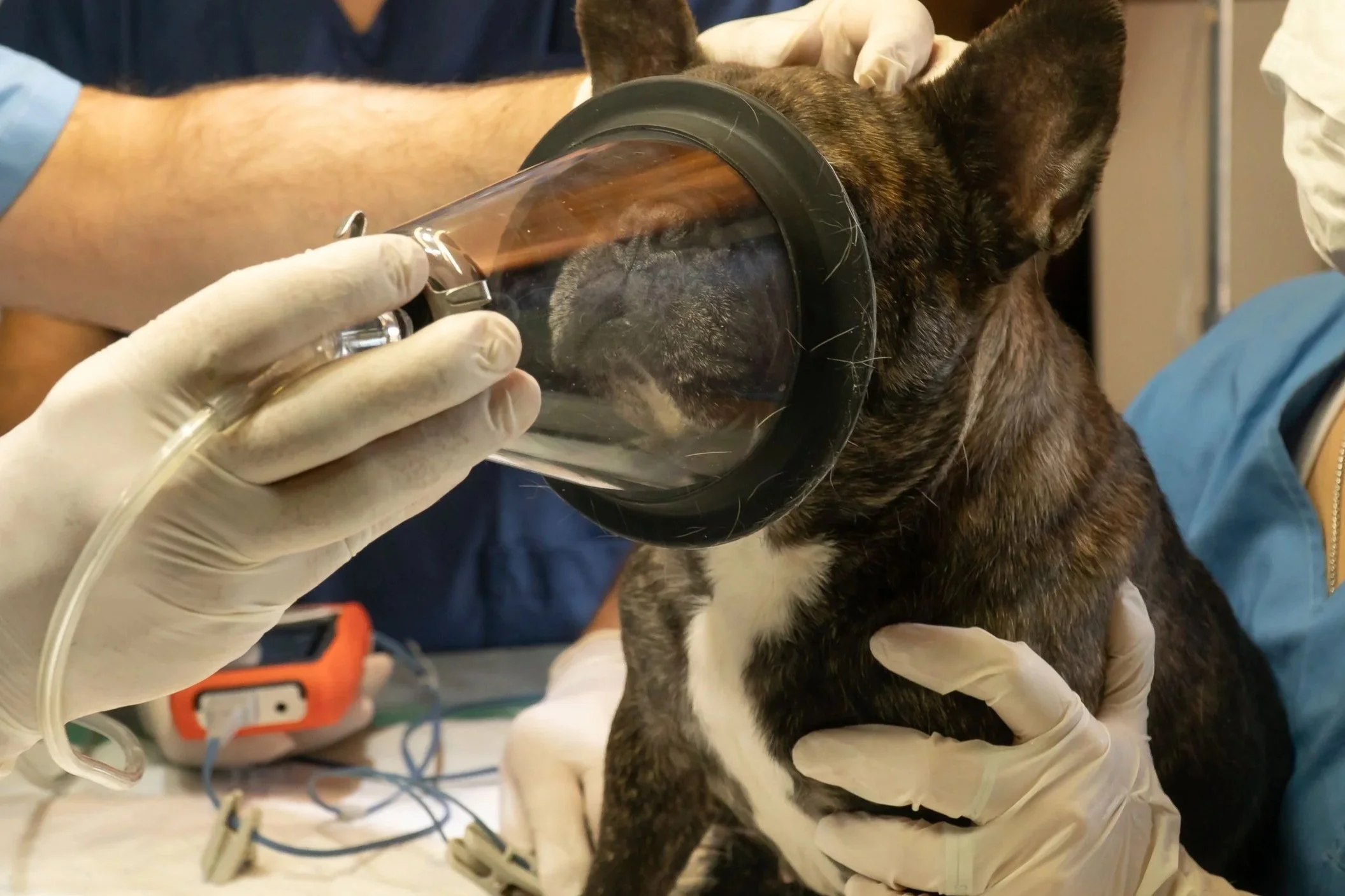
Brachycephalic obstructive airway syndrome can involve stenotic nares, an elongated or thickened soft palate, everted laryngeal tissue, tracheal hypoplasia, and varying degrees of laryngeal collapse. These patients may also have gastroesophageal reflux or regurgitation risk. Stress, sedation, positioning, airway swelling, and loss of pharyngeal muscle tone can all worsen airflow.
A retrospective matched cohort study found that brachycephalic dogs had a higher risk of peri- and postanesthetic complications than nonbrachycephalic dogs, with postanesthetic complications recorded in 13.9% of brachycephalic dogs compared with 3.6% of matched nonbrachycephalic dogs in that study population.
That finding reinforces several practical principles:
Reduce avoidable stress
Agitation increases oxygen demand and can worsen dynamic upper-airway obstruction. A calm environment and an intentional premedication plan can be important parts of airway management.
Prepare for a controlled airway
Preoxygenation is often useful. IV access should be secured as early as practical. The team should have an appropriate laryngoscope and multiple endotracheal tube sizes immediately available because the tracheal lumen may be smaller than expected.
Induction should be organized so that airway control can be established efficiently, with a backup plan for difficult intubation.
Monitor ventilation and oxygenation
Pulse oximetry and capnography are central. Capnography is particularly helpful because a patient receiving supplemental oxygen may maintain oxygen saturation for a period even as ventilation deteriorates.
Treat recovery as a high-risk phase
These patients may obstruct after extubation because of edema, residual anesthetic effect, airway anatomy, pain, or poor positioning. Extubation should be deliberate rather than rushed. Reintubation equipment and personnel should remain immediately available, and oxygen supplementation and continuous observation may need to continue well after the procedure.
In brachycephalic anesthesia, “the surgery is over” should never be confused with “the risk is over.”
Chronic Kidney Disease: Protect Perfusion and Avoid Automatic Fluid Decisions
Patients with chronic kidney disease may have reduced renal reserve even when they appear clinically stable.
Important preanesthetic questions include:
- Is the patient dehydrated?
- Is anemia present?
- Are potassium, sodium, acid-base status, or other electrolytes abnormal?
- What is the patient’s baseline blood pressure?
- Is urine production normal?
- Are nephrotoxic medications or concurrent diseases relevant?
- Can the procedure be shortened or staged?
The kidney depends on adequate perfusion. Hypotension, dehydration, blood loss, and excessive anesthetic depth can all threaten renal blood flow. The response, however, should not be to give every renal patient a high fluid rate.
Fluid therapy should be individualized. A fluid bolus is most useful when a patient is likely to be preload responsive; it is not a universal treatment for hypotension. A patient with cardiac disease, low albumin, or limited ability to handle sodium and water can be harmed by indiscriminate volume administration.
Anesthetic management should focus on maintaining circulation, avoiding prolonged hypotension, correcting dehydration before induction when possible, monitoring blood pressure closely, minimizing unnecessary anesthetic duration, and avoiding excessive hemodilution in anemic patients.
Hepatic Disease: Think Beyond “Drug Metabolism”
The liver is involved in drug metabolism, protein synthesis, glucose regulation, and coagulation. Liver disease can therefore affect anesthesia through several pathways at once.
A patient with significant hepatic dysfunction may have:
- reduced drug clearance,
- hypoalbuminemia,
- altered volume of distribution,
- coagulation abnormalities,
- hypoglycemia,
- portal hypertension or ascites,
- encephalopathy,
- concurrent anemia or systemic illness.
The anesthetic strategy should account for the severity and functional consequence of the disease rather than the presence of an elevated liver enzyme alone.
In general, shorter-acting or titratable agents and lower effective doses may be useful when drug clearance is impaired. Reversible drugs can be advantageous in selected patients. Glucose and coagulation status may require specific attention, and excessive hypotension should be avoided because hepatic blood flow is pressure dependent.
Importantly, not every patient with abnormal liver enzymes has clinically important hepatic insufficiency. The preanesthetic assessment should distinguish biochemical abnormalities from functional failure whenever possible.
Endocrine Disease: Stability Matters More Than the Diagnostic Label
Endocrine patients can be stable elective candidates or profoundly unstable emergency patients.
Diabetes mellitus
For diabetic dogs and cats, the anesthetic plan must integrate fasting, insulin or other glucose-lowering therapy, expected procedure time, blood glucose monitoring, hydration, and the risk of hypoglycemia or hyperglycemia. Poorly controlled diabetes, ketoacidosis, severe dehydration, or significant electrolyte disturbance changes the risk substantially.
Medication decisions should be made for the individual patient rather than copied from a generic internet protocol.
Hyperthyroidism
Uncontrolled hyperthyroidism, especially in cats, can produce tachycardia, hypertension, increased myocardial oxygen demand, arrhythmias, and a hypermetabolic state. Elective anesthesia is generally better undertaken after the patient has been evaluated and appropriately stabilized rather than while severe clinical signs remain uncontrolled.
Hypothyroidism
Clinically significant, poorly regulated hypothyroidism can be associated with bradycardia, impaired myocardial performance, lower metabolic rate, hypothermia, obesity, and slower drug clearance. Concurrent laryngeal paralysis or megaesophagus can further increase airway and aspiration risk in some dogs.
The presence of a low total T4 concentration alone does not necessarily establish true hypothyroidism, because nonthyroidal illness and some medications can suppress thyroid hormone measurements. That distinction should be resolved clinically rather than assumed for anesthetic planning.
Hyperadrenocorticism and other endocrine disease
Patients with hyperadrenocorticism may have hypertension, hypercoagulability, diabetes, muscle weakness, infection risk, and altered wound healing. As with other endocrine disorders, the important question is not simply whether the diagnosis exists but whether clinically important consequences are controlled.
Geriatric Patients: Age Is Not a Disease, but Reserve Changes
Senior dogs and cats frequently require anesthesia for procedures that can meaningfully improve quality of life, particularly dentistry, mass removal, imaging, and orthopedic or pain-related interventions.
Chronologic age alone is not a contraindication to anesthesia. AAHA’s senior care guidance explicitly reinforces the concept that old age itself is not a disease.
What changes with age is physiologic reserve.
Older animals are more likely to have decreased cardiovascular compliance, valvular disease, reduced renal reserve, altered hepatic drug metabolism, respiratory changes, impaired thermoregulation, reduced muscle mass, arthritis, and occult disease. They may be less able to compensate for hypotension, hypoventilation, dehydration, hypothermia, or excessive anesthetic depth.
The practical consequences are straightforward:
- obtain a meaningful history and examination,
- choose diagnostics based on findings rather than age alone,
- stabilize correctable disease,
- titrate anesthetic drugs to effect,
- use balanced analgesia to reduce unnecessary anesthetic depth,
- monitor trends closely,
- support temperature, ventilation, and perfusion,
- expect that recovery may require more time and nursing care.
A review of geriatric veterinary anesthesia emphasizes that appropriate screening, judicious dosing, vigilant monitoring, and supportive care are more important than identifying one “best” anesthetic agent.
Neurologic Disease: Protect the Brain by Protecting Physiology
Patients with intracranial disease, traumatic brain injury, seizures, or suspected increased intracranial pressure require deliberate control of oxygenation, ventilation, blood pressure, and stimulation.
The brain is sensitive to both hypoxemia and inadequate perfusion. Marked hypercapnia can increase cerebral blood flow and may worsen intracranial pressure in susceptible patients, while hypotension can reduce cerebral perfusion.
Patients with seizure disorders should be assessed for seizure frequency, current antiepileptic therapy, metabolic contributors, and evidence of intracranial disease. Perioperative interruption of essential anticonvulsant therapy should be avoided unless there is a specific medical reason and an alternative plan.
These are cases in which a quiet induction and recovery environment, carefully controlled ventilation, and a clear emergency plan can be as important as drug selection.
A Previous Anesthetic Complication Is Valuable Clinical Data
A history such as “my pet had a bad reaction to anesthesia” is too nonspecific to guide a plan, but it should never be dismissed.
Obtain the previous anesthetic record whenever possible.
Was the problem hypotension, apnea, airway obstruction, aspiration, prolonged recovery, hypothermia, dysphoria, arrhythmia, difficult intubation, unexpected drug sensitivity, or something unrelated to anesthesia?
The difference matters.
A documented previous airway obstruction should change preparation and recovery planning. A previous prolonged recovery associated with severe hypothermia suggests a different intervention. A transient low blood pressure reading from an incorrectly sized cuff is not equivalent to refractory shock.
Past records allow the team to convert anxiety into useful information.
Urgency, Duration, and Time of Day Can Change Risk
Patient disease is only one component of risk.
Observational studies have associated anesthesia-related mortality with urgent or nonelective procedures and with higher ASA status. AAHA also notes that late-day or after-hours procedures can carry additional risk because staffing, preparation time, fatigue, and recovery resources may differ.
That does not mean an emergency should wait when delay would be dangerous. It means teams should recognize that an emergency patient anesthetized at 2 a.m. has a different risk environment than a stable elective patient scheduled first thing in the morning with a fully staffed hospital.
For complex elective cases, scheduling should create enough time for preparation and supported recovery.
A Seven-Question Framework for High-Risk Cases
Before proceeding, the anesthesia team should be able to answer these questions:
1. What is the dominant physiologic vulnerability?
Airway? Contractility? Diastolic filling? Perfusion? Gas exchange? Glucose? Coagulation? Intracranial pressure? Drug clearance?
2. What can be improved before anesthesia?
Correct dehydration, stabilize congestive heart failure, address severe electrolyte abnormalities, improve glucose control, treat respiratory distress, or obtain additional diagnostics when the result will change management.
3. What anesthetic effects are least tolerable?
For example, substantial tachycardia may be poorly tolerated in hypertrophic cardiomyopathy, while excessive afterload may be undesirable in mitral regurgitation.
4. What monitoring is essential?
At a minimum, follow current small-animal monitoring guidance. Higher-risk patients may justify invasive blood pressure monitoring, blood gases, serial laboratory testing, more advanced ventilation monitoring, or other modalities.
5. What complications are most likely?
Write them down. Prepare the equipment, drugs, personnel, and escalation plan before induction.
6. What does recovery need to look like?
Will the patient need prolonged oxygen, delayed extubation, active warming, repeated blood pressure checks, additional analgesia, overnight monitoring, or a higher-acuity location?
7. Would additional expertise change management?
If the team lacks experience with the disease, does not have the required monitoring or intervention capability, or would benefit from specialist guidance, consider referral, an on-site specialist, or veterinarian-to-specialist teleconsulting when appropriate.
Where a Board-Certified Veterinary Anesthesiologist Can Add Value
The ACVAA defines veterinary anesthesiology as a specialty focused on total perioperative patient care, pain management, cardiopulmonary support, and management of animals undergoing anesthesia or sedation. ACVAA Diplomates complete formal advanced training and specialty certification.
The 2025 ACVAA small-animal monitoring guidelines state that, as an advanced recommendation, a board-certified veterinary anesthesiologist should lead the anesthesia care team whenever possible.
That involvement can take several forms:
- direct care in a specialty or university hospital,
- an anesthesiologist working within a general or specialty practice,
- a traveling anesthesiologist,
- a preoperative specialist consultation,
- real-time professional-to-professional teleconsulting.
A remote anesthesiologist cannot intubate a patient, place an IV catheter, palpate pulses, or administer a drug from another location. Those physical tasks remain with the on-site team. What real-time specialist support can provide is patient-specific planning, interpretation of changing clinical information, troubleshooting, and decision support while the local team performs hands-on care.
Safe Pet Anesthesia uses a teleconsulting model in which veterinary practices submit records for case review, receive a patient-specific anesthetic plan, and connect with an anesthesia specialist by secure live video during the anesthetic event from induction through recovery.
The purpose is not to make a high-risk patient “risk free.” It is to give the treating veterinary team additional anesthesia expertise when that expertise is not physically on staff.
Frequently Asked Questions
What makes a dog or cat “high risk” for anesthesia?
Higher anesthetic risk can result from systemic disease, poor physiologic reserve, airway anatomy, emergency status, extreme body condition, advanced age combined with comorbidities, prior anesthetic complications, or a procedure that places unusual demands on circulation, ventilation, blood loss, temperature, or recovery.
Does a heart murmur mean a pet cannot have anesthesia?
No. A murmur is a physical finding, not an anesthetic diagnosis. The underlying disease, its severity, cardiac function, clinical signs, and planned procedure matter. Some patients require additional cardiac evaluation before an anesthetic plan can be developed.
Is a geriatric pet automatically high risk?
Not solely because of age. Older patients are more likely to have concurrent disease and reduced reserve, so risk assessment should focus on the individual patient rather than a birthday.
Can kidney disease patients be anesthetized?
Many can, but the plan should consider hydration, electrolytes, anemia, baseline blood pressure, renal reserve, medications, and the need to avoid prolonged hypotension or inappropriate fluid therapy.
Why are brachycephalic dogs often considered higher risk?
Their upper-airway anatomy can make obstruction more likely during sedation, induction, and especially recovery. Some are also predisposed to regurgitation. Preparation for airway management and prolonged observation after extubation can be particularly important.
When should a case be referred?
Referral should be considered when the patient needs diagnostics, monitoring, intervention capability, postoperative care, or specialty expertise beyond what the current facility can safely provide. Real-time teleconsulting may provide additional expertise for some cases, but it does not substitute for equipment or hands-on capabilities that are physically required.
Conclusion
High-risk veterinary anesthesia is not about finding a special “safe” drug.
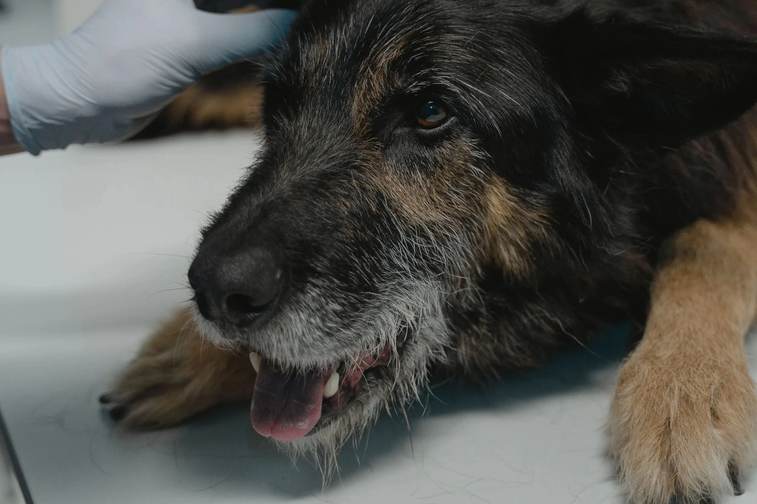
It is about identifying which physiologic systems are vulnerable and building an anesthetic plan that protects them. Cardiac disease requires lesion-specific hemodynamic goals. Brachycephalic patients require an airway plan that extends well into recovery. Renal and hepatic disease require attention to perfusion, fluid balance, metabolism, and systemic consequences. Endocrine disorders should be stabilized when possible. Geriatric patients need thoughtful assessment and careful titration rather than automatic exclusion from beneficial procedures.
The best high-risk plan is individualized, explicit, and prepared for complications before they happen.
References
Grubb T, Sager J, Gaynor JS, et al. 2020 AAHA Anesthesia and Monitoring Guidelines for Dogs and Cats. Journal of the American Animal Hospital Association. 2020;56(2):59-82. https://www.aaha.org/resources/2020-aaha-anesthesia-and-monitoring-guidelines-for-dogs-and-cats/
Bailey K, Briley J, Duffee L, et al. The American College of Veterinary Anesthesia and Analgesia Small Animal Anesthesia and Sedation Monitoring Guidelines 2025. Veterinary Anaesthesia and Analgesia. 2025;52(4):377-385. https://doi.org/10.1016/j.vaa.2025.03.015
Portier K, Ida KK. The ASA Physical Status Classification: What Is the Evidence for Recommending Its Use in Veterinary Anesthesia? A Systematic Review. Frontiers in Veterinary Science. 2018. https://pubmed.ncbi.nlm.nih.gov/30234133/
Pre-anaesthetic risk assessment and management of dogs with myxomatous mitral valve disease: a spectrum of care narrative review. Journal of Small Animal Practice. 2026. https://pubmed.ncbi.nlm.nih.gov/41863286/
Gruenheid M, Aarnes TK, McLoughlin MA, et al. Risk of anesthesia-related complications in brachycephalic dogs. Journal of the American Veterinary Medical Association. 2018;253(3):301-306. https://pubmed.ncbi.nlm.nih.gov/30020004/
Dhaliwal R, Boynton E, Carrera-Justiz S, et al. 2023 AAHA Senior Care Guidelines for Dogs and Cats. Journal of the American Animal Hospital Association. 2023;59(1):1-21. https://pubmed.ncbi.nlm.nih.gov/36584321/
Carpenter RE, Pettifer GR, Tranquilli WJ. Anesthesia for geriatric patients. Veterinary Clinics of North America: Small Animal Practice. 2005;35(3):571-580. https://pubmed.ncbi.nlm.nih.gov/15833559/
Matthews NS, Mohn TJ, Yang M, et al. Factors associated with anesthetic-related death in dogs and cats in primary care veterinary hospitals. Journal of the American Veterinary Medical Association. 2017;250(6):655-665. https://pubmed.ncbi.nlm.nih.gov/28263113/
American College of Veterinary Anesthesia and Analgesia. Residency Training Standards: Definition and Scope of Discipline. https://acvaa.org/wp-content/uploads/2020/06/ACVAA_Residency_Training_Standards_2017.websiteT-July-2020-No-LINK-clean-.pdf
dvm360. Introducing Safe Pet Anesthesia. November 5, 2024. https://www.dvm360.com/view/introducing-safe-pet-anesthesia
dvm360. Transforming veterinary anesthesia with teleconsulting. March 10, 2026. https://www.dvm360.com/view/transforming-veterinary-anesthesia-with-teleconsulting
Continue Reading