Veterinary Anesthesia Safety: A Practical Framework From Preanesthetic Assessment Through Recovery"
Gianluca Bini, DVM MRCVS DACVAA
Anesthesia safety is not the absence of risk. It is the deliberate management of risk before, during, and after an anesthetic event.
That distinction matters because there is no single drug, monitor, or protocol that makes anesthesia “safe” by itself. A stable outcome is usually the product of a system: appropriate patient assessment, a patient-specific plan, a trained team, functioning equipment, careful airway management, effective analgesia, continuous physiologic monitoring, timely intervention, and attentive recovery care.
For veterinary teams, the most useful question is therefore not, “What is the safest anesthetic drug?” It is, “What risks does this patient bring to this procedure, and how will our team recognize and respond to problems throughout the entire anesthetic period?”
Current guidance supports this systems approach. The 2020 American Animal Hospital Association (AAHA) anesthesia guidelines describe anesthesia as a continuum that begins before induction and extends through recovery and return home. The 2025 American College of Veterinary Anesthesia and Analgesia (ACVAA) monitoring guidelines similarly emphasize dedicated anesthesia personnel, hands-on assessment, electronic monitoring, communication, and cognitive aids such as checklists.
This article reviews the elements of a practical anesthesia-safety framework for dogs and cats and explains where additional specialist support may fit.
1. Start With the Patient, Not the Drug
The foundation of safer anesthesia is a meaningful preanesthetic assessment.
A complete evaluation should identify conditions that could change the anesthetic plan, including cardiovascular disease, respiratory compromise, renal or hepatic dysfunction, endocrine disease, anemia, dehydration, electrolyte abnormalities, neurologic disease, obesity or cachexia, age-related physiologic changes, previous anesthetic complications, and medications or supplements that may alter the response to anesthesia.
The history is not a formality. Exercise intolerance, coughing, collapse, vomiting or regurgitation, changes in water intake, weight loss, altered mentation, recent illness, and a difficult previous recovery can all be clinically relevant. The physical examination should be current and should pay particular attention to airway anatomy, respiratory effort, heart rate and rhythm, perfusion, hydration, body condition, temperature, and any findings related to the planned procedure.
AAHA recommends using the American Society of Anesthesiologists physical status classification as one framework for summarizing patient health. It is not a complete risk calculator, but higher ASA status has repeatedly been associated with greater anesthesia-related mortality in veterinary patients.
Preanesthetic testing should be selected for the individual patient rather than ordered reflexively. A healthy young patient and a geriatric dog with a murmur, weight loss, and chronic kidney disease do not need identical diagnostic workups. The goal is to obtain information that can identify disease, alter the anesthetic plan, prompt stabilization, or change the timing or location of the procedure.
Stabilization is part of anesthesia
Sometimes the safest anesthetic decision is to delay an elective procedure.
AAHA specifically identifies abnormalities such as dehydration, significant anemia, hypoglycemia, hypothermia, life-threatening arrhythmias, major electrolyte or acid-base disturbances, congestive heart failure, cyanosis, and severe urinary abnormalities as conditions that may require correction or stabilization before anesthesia.
“Proceed or postpone?” is therefore a clinical decision, not a scheduling decision. If a correctable abnormality can meaningfully reduce risk, addressing it before an elective anesthetic event may be more important than changing the induction drug.
2. Build an Individualized Plan for the Patient, Procedure, and Practice
A good anesthetic plan is sequential. It considers what should happen before premedication, during induction, during maintenance, at the end of the procedure, during extubation, and throughout recovery.
The plan should account for three variables at the same time:
The patient. What physiologic reserve does this animal have? What organ systems are vulnerable? What adverse effects would be poorly tolerated?
The procedure. How painful will it be? How long is it likely to take? Will positioning affect ventilation or venous return? Will the airway be accessible? Is significant blood loss possible? Is the procedure elective or urgent?
The practice environment. Which drugs, monitoring modalities, oxygen-delivery systems, ventilators, warming devices, emergency medications, staff members, and advanced diagnostics are actually available?
This is why “recipe anesthesia” has limits. A protocol that is appropriate for a young healthy dog undergoing a short elective procedure may be a poor fit for a geriatric patient with mitral valve disease, a cat with hypertrophic cardiomyopathy, or a brachycephalic dog with upper-airway obstruction.
Balanced anesthesia can also reduce reliance on any one anesthetic component. Appropriate premedication, analgesia, locoregional techniques, and carefully titrated induction and maintenance agents may allow lower doses of drugs that cause dose-dependent cardiovascular or respiratory depression. The objective is not to use the fewest medications. It is to use each component intentionally.
3. Treat the Anesthesia Team as a Safety System
Equipment matters, but people detect context.
The 2025 ACVAA guidelines list a dedicated anesthetist as a minimum recommendation for anesthetized small animals. The dedicated person should not simply record values. That individual must continuously integrate physical examination findings, anesthetic depth, the monitor display, trends over time, procedural events, and the patient’s response to interventions.
This distinction is critical. A multiparameter monitor can show a blood pressure, heart rate, oxygen saturation, and end-tidal carbon dioxide value. It cannot independently determine whether a change is caused by excessive anesthetic depth, inadequate ventilation, blood loss, poor venous return, equipment error, airway obstruction, a disconnected IV catheter, or surgical stimulation.
Anesthesia safety depends on interpretation.
Checklists reduce reliance on memory
Checklists and cognitive aids are not substitutes for knowledge. They protect teams from predictable human limitations.
A useful anesthesia checklist may confirm patient identity, procedure, fasting status, allergies, recent medications, airway equipment, endotracheal tube sizes, IV access, anesthetic machine leak testing, oxygen supply, monitoring equipment, emergency drugs, analgesic plan, fluid plan, warming strategy, anticipated complications, and recovery responsibilities.
A short team briefing can also clarify who is monitoring, who is performing the procedure, who will respond if an emergency occurs, and what the escalation plan is.
The 2025 ACVAA guidance recommends direct and frequent communication between the anesthetist and the veterinary team and supports surgical safety checklists tailored to the clinical environment.
4. Monitor the Patient, Not Just the Monitor
The 2025 ACVAA small-animal guidelines identify minimum monitoring recommendations that include physical assessment of anesthetic depth, continuous ECG, noninvasive blood pressure measurement, pulse oximetry, capnography, and temperature measurement, together with a dedicated anesthetist.
Each modality answers a different question.
ECG: What is the electrical rhythm?
ECG monitoring helps identify heart rate and rhythm changes. It does not tell you whether the rhythm is producing adequate blood flow. An apparently acceptable ECG can coexist with severe hypotension.
Blood pressure: Is perfusion pressure adequate?
Blood pressure is an imperfect surrogate for tissue perfusion, but it is clinically useful and actionable. Persistent hypotension should prompt assessment of anesthetic depth, heart rate, preload, contractility, vascular tone, blood loss, and measurement accuracy.
A single low number is less informative than a confirmed trend plus the clinical context.
Pulse oximetry: Is hemoglobin adequately saturated with oxygen?
Pulse oximetry helps detect hypoxemia, but interpretation should include waveform or signal quality, perfusion, airway status, inspired oxygen, and ventilation. A patient receiving supplemental oxygen can maintain a normal SpO2 for a period despite clinically important hypoventilation.
Capnography: Is the patient ventilating, and is the airway connected?
Capnography provides continuous information about exhaled carbon dioxide and the shape of the capnogram. It can help identify hypoventilation, apnea, circuit disconnection, airway obstruction, rebreathing, and changes in perfusion. After intubation, detection of exhaled carbon dioxide is also an important method of confirming tracheal placement of the endotracheal tube.
Temperature: Is the patient maintaining thermal homeostasis?
Anesthesia impairs thermoregulation and commonly promotes heat loss. Hypothermia can alter drug handling, delay recovery, increase oxygen demand during shivering, and complicate interpretation or treatment of other physiologic abnormalities. Active warming and minimizing unnecessary exposure should be planned rather than added only after the patient is already cold.
Anesthetic depth: What does the patient look like?
Eye position, palpebral response, jaw tone, muscle tone, spontaneous movement, cardiovascular response to stimulation, and other physical findings remain essential. Electronic monitoring complements hands-on assessment; it does not replace it.
5. Recognize Common Complications Early and Think Mechanistically
Many anesthetic complications are predictable consequences of anesthetic drugs, patient disease, positioning, or the procedure itself. Early recognition makes them more manageable.
Hypotension
When blood pressure falls, ask why.
Mean arterial pressure is influenced by cardiac output and systemic vascular resistance. Cardiac output depends on heart rate and stroke volume, which in turn is influenced by preload, contractility, and afterload. A useful response to hypotension therefore begins by confirming the measurement and identifying the likely mechanism rather than automatically giving the same treatment to every patient.
Potential contributors include excessive anesthetic depth, bradycardia, vasodilation, reduced venous return, dehydration, hemorrhage, impaired myocardial contractility, arrhythmia, or a combination of factors.
Hypoventilation and hypoxemia
General anesthetics commonly depress ventilation. Airway obstruction, bronchial intubation, equipment problems, atelectasis, pulmonary disease, and low cardiac output can also affect gas exchange.
This is why oxygen supplementation alone is not a complete respiratory strategy. A patient can receive a high inspired oxygen concentration and still retain carbon dioxide because ventilation is inadequate. Capnography and pulse oximetry provide complementary information.
Bradycardia and arrhythmias
Heart rate should be interpreted in relation to species, patient baseline, anesthetic depth, blood pressure, perfusion, and the rhythm itself. Not every bradycardic patient requires the same response, and not every arrhythmia is equally important. Treatment should be driven by hemodynamic consequence and underlying mechanism.
Hypothermia
Small patients, geriatric animals, long procedures, open body cavities, cool environments, and large exposed surface areas can increase heat loss. Prevention is often easier than correction.
6. Airway Management Is a Core Safety Skill
For general anesthesia, securing and managing the airway is often one of the most consequential tasks the team performs.
The team should select appropriate endotracheal tube sizes in advance and have alternatives immediately available. Tube placement should be verified, the cuff managed appropriately, and the breathing circuit connected correctly. Capnography can help confirm tracheal placement and provide ongoing evidence that ventilation is reaching the sampling site.
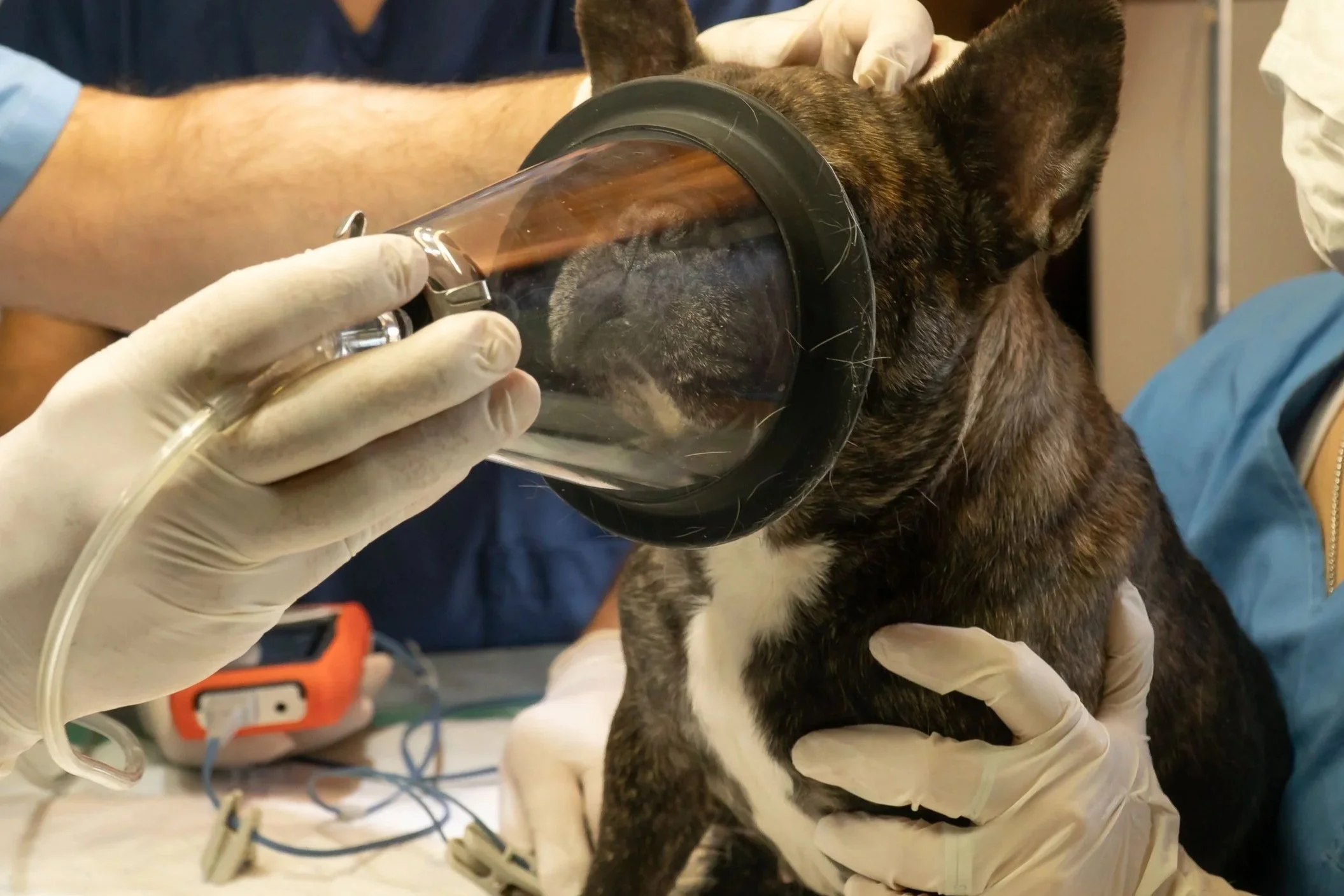
Airway plans need to be patient specific. Brachycephalic dogs, cats prone to laryngospasm, patients at risk of regurgitation, animals with laryngeal disease, and patients undergoing oral or airway surgery may require different induction, intubation, extubation, and recovery strategies.
The key is anticipation. Airway emergencies are safer to manage when the equipment, drugs, personnel, and next step have been considered before induction.
7. Analgesia Is Part of Anesthetic Safety
Pain is not only a welfare concern. Nociception can contribute to sympathetic stimulation, movement, unstable anesthetic depth, and higher maintenance-anesthetic requirements.
A preventive, multimodal analgesic plan may include systemic analgesics and local or regional anesthesia when appropriate. In dentistry, for example, AAHA notes that local anesthetic dental blocks can reduce inhalant requirements and support blood pressure and ventilation while improving analgesia.
Good analgesia can also improve recovery quality. A quiet patient is not automatically a comfortable patient, so structured pain assessment should continue after anesthesia.
8. Recovery Is Not an Afterthought
One of the most important safety lessons in veterinary anesthesia is that the end of the procedure is not the end of anesthetic risk.
A large prospective worldwide study of 55,022 dogs reported that 81% of anesthesia-related deaths occurred postoperatively. A separate multicenter study of 14,962 cats reported that 74.5% of anesthesia-related deaths occurred postoperatively. These studies included diverse patients and procedures, so their overall mortality rates should not be used as a prediction for an individual animal. The distribution of deaths, however, reinforces a practical point: recovery deserves the same deliberate planning as induction and maintenance.
Recovery should include continued physical observation and monitoring appropriate to the patient until physiologic function and airway control are reliable.
Questions to consider include:
- Is the patient ventilating adequately?
- Is oxygen saturation acceptable?
- Is body temperature improving?
- Is blood pressure or perfusion still a concern?
- Is pain controlled?
- Is nausea or regurgitation a risk?
- Is the patient able to maintain and protect the airway?
- Is dysphoria being distinguished from pain or hypoxemia?
- Does this patient need prolonged oxygen supplementation or a higher level of postanesthetic observation?
Handoffs are especially important. If the anesthetist transfers care to another team member, the receiving person should know the anesthetic protocol, complications, treatments given, outstanding concerns, analgesic plan, and discharge criteria.
9. Some Patients Need a Different Level of Planning
“High risk” is not a diagnosis. It is a description of the relationship between the patient’s physiologic reserve, the planned procedure, and the resources available to manage complications.
Cases that may justify additional planning or specialist input include patients with significant cardiac disease, brachycephalic airway disease, severe pulmonary disease, advanced renal or hepatic dysfunction, unstable endocrine disease, major anemia, previous serious anesthetic complications, neurologic disease affecting ventilation or intracranial pressure, severe obesity or cachexia, and some geriatric patients with multiple comorbidities.
The 2025 ACVAA guidelines state as an advanced recommendation that a board-certified veterinary anesthesiologist should lead the anesthesia care team whenever possible.
That leadership can be available in different ways depending on the hospital: an in-house anesthesiologist, referral to a specialty or academic center, a traveling anesthesiologist, or veterinarian-to-specialist teleconsulting where legally and clinically appropriate.
10. Where Real-Time Veterinary Anesthesia Teleconsulting Fits
Teleconsulting does not replace the veterinarian and veterinary team physically caring for the patient. It can provide an additional specialist perspective to the on-site team.
In a live anesthesia teleconsulting model, the specialist may review the medical record and diagnostics before the case, help develop a patient-specific plan, then remain available by live video during induction, maintenance, and recovery. The practice team remains responsible for hands-on patient care, drug administration, airway management, monitoring connections, interventions, and compliance with applicable veterinary practice requirements.
Safe Pet Anesthesia uses this model: veterinary practices submit patient information for review, receive an individualized protocol, and connect with an anesthesia team by secure live video from induction through recovery. Independent veterinary-industry reporting by dvm360 has also described this workflow.
Teleconsulting should be viewed as expert support, not as a guarantee against complications. Safe anesthesia still depends on an adequately trained on-site team, appropriate equipment, patient assessment, monitoring, communication, and the ability to intervene physically when needed.
A Practical Veterinary Anesthesia Safety Checklist
Before induction, ask:
1. What are this patient’s specific risk factors?
2. Does anything need to be stabilized before proceeding?
3. Is the protocol individualized to the patient and procedure?
4. Is a dedicated team member assigned to anesthesia?
5. Are the anesthesia machine, airway equipment, monitors, warming equipment, IV access, and emergency drugs ready?
6. What complications are most likely, and what is the response plan?
7. What is the analgesic strategy?
8. What are the extubation and recovery criteria?
9. Who owns the patient during recovery and handoff?
10. Would specialist involvement meaningfully improve planning or real-time decision support for this case?
Frequently Asked Questions
Is veterinary anesthesia safe?
No anesthetic event is risk free. Risk varies substantially with patient health, species, age, body condition, urgency, procedure, team experience, and the care environment. The goal is not to promise zero risk but to identify and reduce modifiable risks through assessment, individualized planning, monitoring, physiologic support, and recovery care.
What monitoring should an anesthetized dog or cat have?
The 2025 ACVAA guidelines list a dedicated anesthetist, physical assessment of anesthetic depth, continuous ECG, noninvasive blood pressure, pulse oximetry, capnography, and temperature measurement among minimum recommendations for anesthetized small animals. Individual patients may require additional monitoring.
Is an older pet automatically too risky for anesthesia?
No. Age is not a disease. Older animals are more likely to have reduced physiologic reserve or concurrent disease, so they may require more thorough assessment, dose titration, supportive care, and recovery monitoring. The decision should be based on the individual patient and the benefit of the planned procedure.
Why is recovery monitoring so important?
Respiratory depression, airway obstruction, hypothermia, pain, dysphoria, regurgitation, and hemodynamic problems can persist or emerge after anesthetic drugs are reduced or discontinued. Recent large observational studies in dogs and cats found that most anesthesia-related deaths occurred after the procedure rather than during maintenance.
Can a general practice involve a board-certified veterinary anesthesiologist?
Options may include referral, traveling specialist services, or professional-to-professional tele-consulting. The best model depends on the patient, procedure, local regulations, practice capabilities, and availability.
Conclusion
Veterinary anesthesia safety is a process. It begins with accurate assessment and honest risk recognition. It continues with a patient-specific plan, a prepared team, appropriate analgesia, airway management, hands-on observation, and continuous physiologic monitoring. It requires a mechanism-based response when complications occur. And it continues through recovery until the patient is physiologically stable and able to protect its airway.
There is no magic drug combination that can replace those fundamentals.
For veterinary practices that want additional anesthesia expertise for selected or routine cases, board-certified specialist support can be incorporated through referral, on-site services, or real-time teleconsulting. The purpose is not to replace the team at the patient’s side. It is to give that team more expertise to draw on while they provide the hands-on care.
References
Grubb T, Sager J, Gaynor JS, et al. 2020 AAHA Anesthesia and Monitoring Guidelines for Dogs and Cats. Journal of the American Animal Hospital Association. 2020;56(2):59-82. https://www.aaha.org/resources/2020-aaha-anesthesia-and-monitoring-guidelines-for-dogs-and-cats/
Bailey K, Briley J, Duffee L, et al. The American College of Veterinary Anesthesia and Analgesia Small Animal Anesthesia and Sedation Monitoring Guidelines 2025. Veterinary Anaesthesia and Analgesia. 2025;52(4):377-385. https://doi.org/10.1016/j.vaa.2025.03.015
Portier K, Ida KK. The ASA Physical Status Classification: What Is the Evidence for Recommending Its Use in Veterinary Anesthesia? A Systematic Review. Frontiers in Veterinary Science. 2018. https://pubmed.ncbi.nlm.nih.gov/30234133/
Bellows J, Berg ML, Dennis S, et al. 2019 AAHA Dental Care Guidelines for Dogs and Cats. Journal of the American Animal Hospital Association. 2019;55(2):49-69. https://www.aaha.org/resources/2019-aaha-dental-care-guidelines-for-dogs-and-cats/
Redondo JI, Otero PE, Martínez-Taboada F, et al. Anaesthetic mortality in dogs: A worldwide analysis and risk assessment. Veterinary Record. 2024;195(1):e3604. https://pubmed.ncbi.nlm.nih.gov/37953683/
Redondo JI, Martínez-Taboada F, Viscasillas J, et al. Anaesthetic mortality in cats: A worldwide analysis and risk assessment. Veterinary Record. 2024;195(1):e4147. https://pubmed.ncbi.nlm.nih.gov/38959210/
dvm360. Introducing Safe Pet Anesthesia. November 5, 2024. https://www.dvm360.com/view/introducing-safe-pet-anesthesia
dvm360. Transforming veterinary anesthesia with teleconsulting. March 10, 2026. https://www.dvm360.com/view/transforming-veterinary-anesthesia-with-teleconsulting
Continue Reading